
Di seguito alcune immagini che possono facilitare la comprensione di alcune strutture che abbiamo trattato in classe 5E BTS: 
 |
| Corpo ciliare e pupilla |
 |
| Iride |
 |
Vertical section of a chick retina
(400x) |
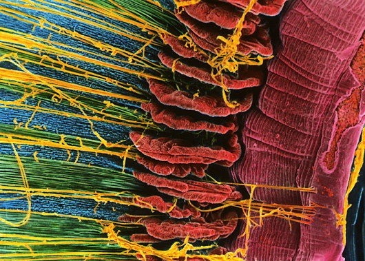
![Retina. Coloured scanning electron micrograph (SEM) of rods (yellow) and cones (green) in the retina of the eye. The outer nuclear layer is purple. Magnification x1800 when printed at 10 centimetres wide. [F0010041] Incredible!!:](https://s-media-cache-ak0.pinimg.com/564x/05/85/3e/05853e663cb055396b935cffd984365c.jpg) |
| Coni (verdi) e Bastoncelli (grigi). Lo strato esterno nucleare è in viola. SEM x1800 |
 |
| Coni e Bastoncelli della retina . Microscopio a scansione elettronica. |
 |
| Come gli occhi vedono a colori |
 |
| Aggiungi didascalia |
Commenti
Posta un commento